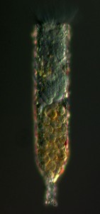
Of the approximately 2,000 known species of living dinoflagellates, about 150 are parasitic. These organisms can alter the marine food web, in some cases destroying prey that consumers like copepods and larval fish rely upon. Coats first spotted T. acutus in the 1980s, in plankton samples he had collected from the Chesapeake Bay. Through his microscope, he noticed a ciliate being edged out of its lorica (shell) by a dinoflagellate. It looked different from others he had observed.

The host lorica (shell) contains the host ciliate Tintinnopsis cylindrica (upper part), which is being consumed by the parasitic dinoflagellate Tintinnophagus acutus (bottom, yellow). Photo: Wayne Coats
Much of Coats’ work involved understanding, questioning and clarifying various accounts of similar dinoflagellates that have been written over the years. He read studies published in French, German and English. This thorough research resulted in more than the introduction of T. acutus: it provided new understanding of the evolutionary relationships among parasitic dinoflagellates and it better defined their position within the dinoflagellate lineage of the tree of life.
Protist phylogeny has never been Coats’ primary focus. T. acutus is the second species that he has named and described. This fall Coats will retire from SERC; he says he expects to have time to describe a few more species of parasitic dinoflagellates.
Tintinnophagus acutus, Just the Facts
Translation: Pointed tintinnid eater.

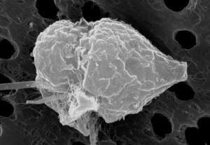
A scanning electron microscopy image of a Tintinnophagus acutus dinospore. The dinospore attaches to the outside of the host with its peduncle (feeding tube). It will feed, grow and divide over the next 4 to 6 days and ultimately produce a number of infective dinospores that will go in search of a host. Image: Wayne Coats
Host Organism
T. acutus is known to parasitize Tintinnopsis cylindrica, a ciliate found in the Chesapeake Bay during the winter months.
Ecto or Endo?
T. acutus is an ectoparasite; it attaches its peduncle (feeding tube) to the outside of its host to feed. This distinguishes T. acutus from other dinoflagellates that parasitize ciliates, which are all endoparasites and live inside the host’s cytoplasm.
Impact on Host
Ciliates infected with T. acutus are smaller than uninfected ciliates and appear to lose their ability to reproduce. The host can survive infections, but in some cases the ciliate abandons its lorica (shell), leaving it without any protection. When this happens, the ciliate has to spend extra energy building a new lorica.
Want to read more? “Tintinnophagus acutus n.g., n. sp. (Phylum Dinoflagellata), an Ectoparasite of the Ciliate Tintinnopsis cylindrica Daday 1887, and Its Relationship to Duboscuodinium collini Grassé 1952,” by D. Wayne Coats, Sunju Kim, Tsvetan R. Bachvaroff, Sara M. Handy and Charles F. Delwiche will be published in an upcoming issue of the Journal of Eukaryotic Microbiology.


