by Katrina Lohan
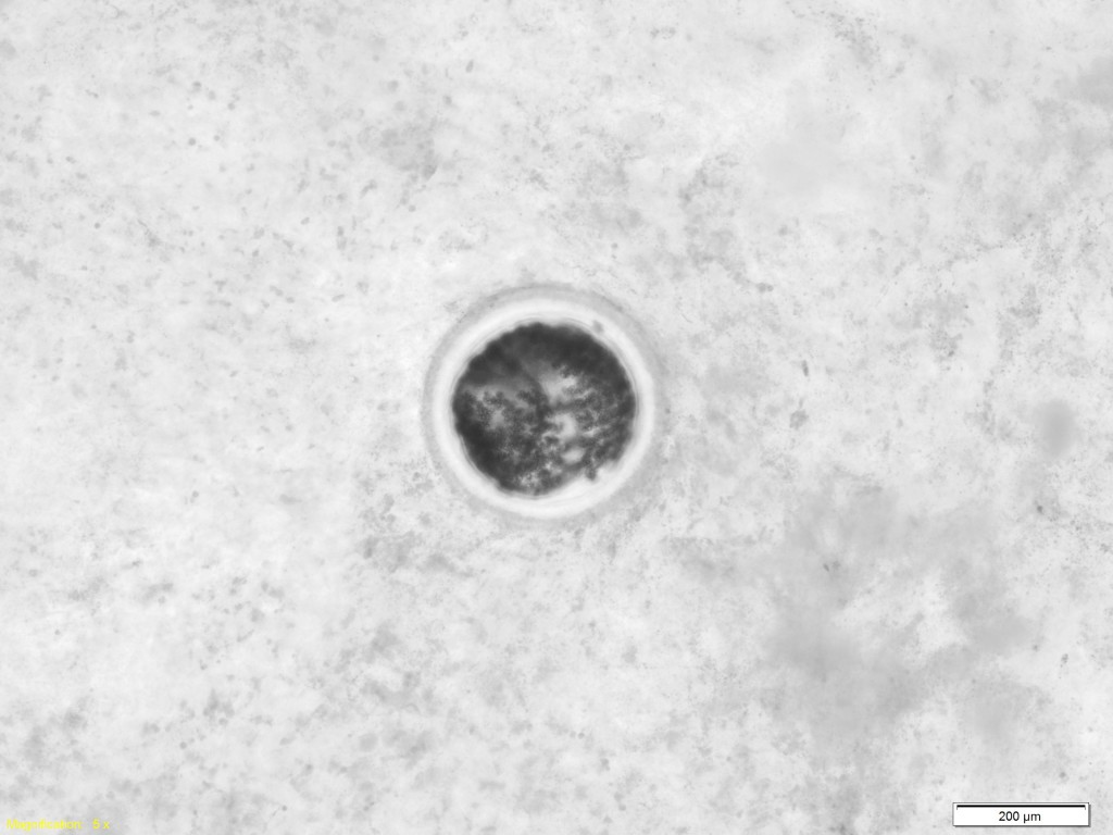
Microscopic slide of a mussel parasite called a trematode. Trematodes often infect small aquatic animals, like mussels or snails, in hope of getting eaten by a larger host they can infect later. Viewed under 400x magnification. (Katrina Lohan)
Just as in previous trips, part of our sampling involved dissecting the bivalves and preparing tissue preps to view under the microscope. I have to admit that the microscopic metazoan parasites that I saw on this trip were not quite as exciting as previous trips—I miss the really cute turbellarians (a.k.a.
Urostoma sp.)! Though they weren’t as cute, I did see some parasites through the microscope, including trematode metacercariae in the mussels, three marine mites, a handful of pea crabs and free-swimming harpacticoid copepods. I had never seen a marine mite before and at first thought there were ticks running around the lab. That’s what happens when you spend hours in front of a microscope–you forget how small the things you are viewing actually are!


